Principal investigator: László Négyessy
The continuous sheet of the cerebral cortex is a complex network of structurally and functionally heterogeneous areas. In higher order mammals including primates, the cortical network is formed by populations of closely spaced neurons with convergent or overlapping afferents within and between the areas. Thereby connections appear modular or patch-like both within and between the areas. Similarly, cortical activations are spatiotemporally delineated forming a distributed modular architecture in the functioning brain. As such, modular organization represents the transition from the micro circuits to the large-scale network of the areas. In some areas the modular connectional architecture corresponds to the known columnar cortical organization, such that certain kinds of columns are selectively connected to each other (most notably the orientation columns of the visual cortex). However, in most of the cases connectional preferences of the neuronal populations are not known. A major obstacle in understanding the functioning of the cerebral cortex is that the modular architecture (or in other terms population level, or meso-scale connections) is largely unexplored. We apply theoretical and experimental approaches to expand our knowledge of the meso-scale cortical network of primates. Our findings can be operative in developing novel brain machine interfaces (BMI).
From the perspective of network science
Our studies are focused on the investigation of the anatomical network where the directions of connections are known. We aim to explore and understand unique features determining cortical signal flow via computational simulations and analyses.
Some picks from our findings:
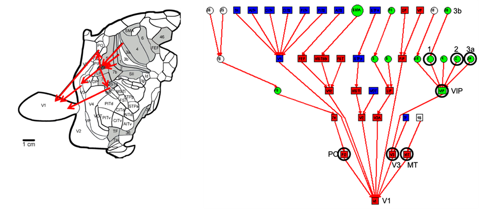
Functional substitution following deafferentation
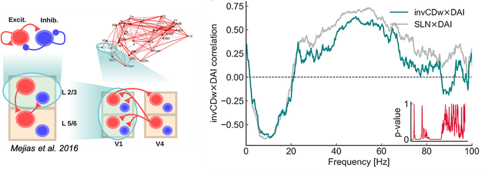
Topological basis of the hierarchical counterstream
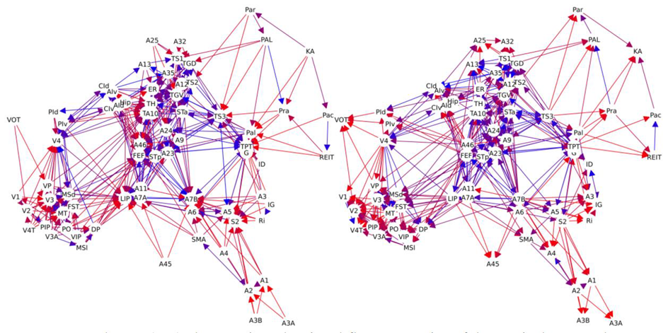
Effects of network topology on its dynamics
Experimental approaches
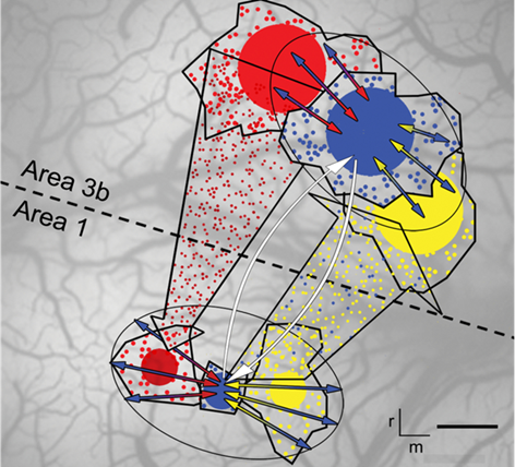
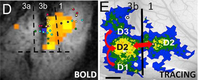
We study the neuronal connectivity of functionally identified modules of the primate somatosensory cortex. In this project we use neuroanatomical tracing aided by tactile stimulation, imaging techniques (intrinsic signal and fMRI) and electrophysiological mapping. We focus on the hand representation region, which is central to tactile functions, and we determine the convergence and divergence of intrinsic, feedforward and feedback connections of neuronal populations. Our aims are twofold: i) to understand the connectional basis of tactile perception and ii) to determine connectional motifs of the meso-scale cortical network.
Some highlights from our findings:
Relationship of anatomical and functional connectivity at the meso-scale
Connectional motif of the columnar circuitry